
Tiny but Powerful Travelers
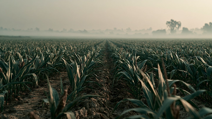
In everyday life, we rarely pay attention to what lurks in the air or dust around us. But under the microscope, you would see countless fungal spores. They are only a few micrometers in diameter, yet they can survive for long periods in harsh environments. These tiny particles are what allow fungi to thrive across the globe, from decaying wood and damp walls to even inside our respiratory tracts.

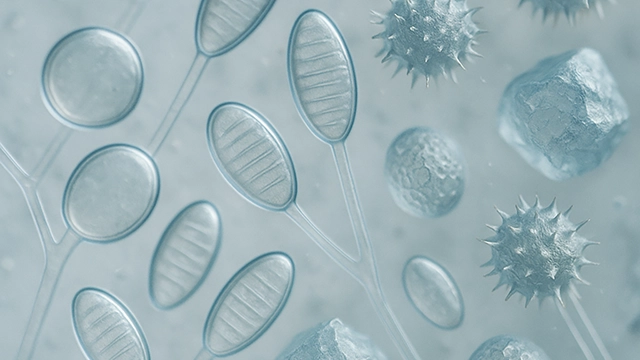
Scanning electron micrograph of fungal spores, Fossil trilete spores (blue) and a spore tetrad (green)
The Ever-Changing Shapes of Spores
Think of the microscope as a telescope, but instead of gazing into distant galaxies, you are observing a “spore universe” that is even more complex. Among the most common are conidia, or asexual spores. Some are perfectly round and smooth, resembling strings of glistening glass beads shining under the light. Others stretch into oval or spindle-like shapes, neatly aligned like miniature grains of rice. Still others are covered with tiny spines and projections, looking like microscopic durians. Their rough surfaces help them stay suspended in the air or cling tightly to surfaces.
Looking further, chlamydospores, or thick-walled spores, resemble warriors clad in armor. Their multiple protective layers allow them to sleep for years in dry soil or nutrient-poor environments. In contrast, melanized spores appear dark and heavy, with coarse textures. Their pigment acts like sunscreen, shielding them from ultraviolet rays and even radiation. Some ascospores, produced inside sac-like structures, are examples of nature’s imagination at work: some coil into spiral springs, others form stars or teardrops, and still others are etched with fine lattice patterns like miniature works of art.
On another front, basidiospores hide quietly beneath the gills of mushrooms, waiting for changes in humidity. When the moment comes, they are catapulted outward like invisible fireworks—releasing billions at once, filling the forest air. If you walk in the woods, you may inhale tens of thousands of such spores with every breath.

There are also lesser-known forms. Microconidia are tiny, almost like floating dust particles, while macroconidiaare large, multi-celled, and intricate like small crystals. Aleurioconidia, which bud directly from hyphae, look plump and ready to detach, spreading like plant seeds. Some fungi produce phragmospores, segmented like pieces of bamboo, each chamber carrying the potential for life. Put together, these countless shapes reveal that fungal spores are not monotonous dust but a vibrant, richly structured microcosm. Each form tells a story—about survival in the environment, about resisting threats, and about finding new places to settle.
The Secrets of Structure and Defense
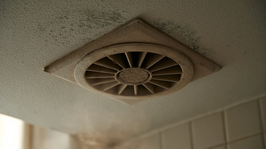
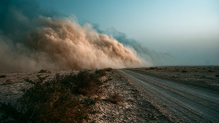
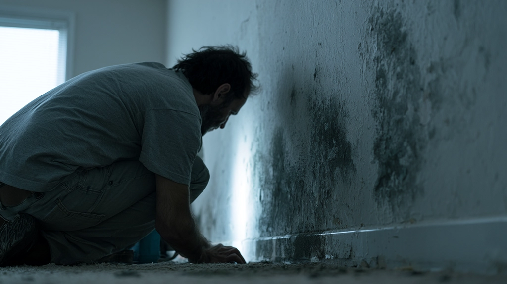
The form and structure of spores largely determine their resilience. Chlamydospores, with their thick cell walls, can withstand dryness and nutrient scarcity, allowing them to persist for long periods in soil or even construction materials. Melanized spores, with their dark pigmentation, are like protective shields against ultraviolet radiation, enabling survival in deserts, polar regions, and even radioactive sites.
The spines and rough ridges on spore surfaces are not random decorations. Research shows they increase friction, making it easier for spores to stick to insect cuticles, animal fur, or even human clothing. Through this “hitchhiking” strategy, fungi can spread quickly into new habitats.

Spores as Taxonomic Fingerprints
To mycologists, spores serve as a fungus’s “identity card.” In classification and identification, features such as spore size, shape, color, surface texture, and arrangement are critical. For example, Aspergillus spores are carried on bottle-shaped structures called conidiophores, radiating outward, while Penicillium spores are produced on branched conidiophores, forming brush-like clusters. These differences are classical diagnostic traits, still essential in food safety, medicine, and building inspections.
In recent years, scientists have combined geometric morphometrics—a method for quantitatively analyzing shapes—with molecular biology techniques to achieve more precise classification. In this sense, spore morphology is not only the result of evolution but also a key to unlocking the fungal world.

Stories of Survival Hidden in Form
These seemingly insignificant particles are actually fungi’s greatest survival weapon. Their forms not only dictate how well they withstand environmental pressures but also influence how efficiently they spread and attach. When we peer at spores through the microscope, we are not just looking at strange geometrical structures—we are witnessing the long history of fungal adaptation and interaction with their surroundings.
So, the next time you clean your home and see dust rise into the air, remember that hidden within may be these “miniature sculptures.” They may be small, but they hold the secrets of how fungi continue to thrive in every corner of the Earth. To read the shape of a spore is to read the survival story of fungi themselves.
References
- Latgé JP & Chamilos G. (2019). Aspergillus fumigatus and aspergillosis in 2019. Clinical Microbiology Reviews, 33(1). DOI:10.1128/CMR.00140-18
- Wyatt TT, Wösten HAB, Dijksterhuis J. (2013). Fungal spores for dispersion in space and time. Advances in Applied Microbiology, 85:43–91. DOI:10.1016/B978-0-12-407672-3.00002-2
- WHO. Fungal Pathogens Priority List. WHO
Key Takeaways
- In everyday life, we rarely pay attention to what lurks in the air or dust around us.
- They are only a few micrometers in diameter, yet they can survive for long periods in harsh environments.
- Some are perfectly round and smooth, resembling strings of glistening glass beads shining under the light.
- Still others are covered with tiny spines and projections, looking like microscopic durians.
- Their multiple protective layers allow them to sleep for years in dry soil or nutrient-poor environments.
Frequently Asked Questions
What should you know about tiny but Powerful Travelers?
In everyday life, we rarely pay attention to what lurks in the air or dust around us. But under the microscope, you would see countless fungal spores. They are only a few micrometers in diameter, yet they can survive for long periods in harsh environments. These tiny particles are what allow fu
What should you know about the Ever-Changing Shapes of Spores?
Think of the microscope as a telescope, but instead of gazing into distant galaxies, you are observing a “spore universe” that is even more complex. Among the most common are conidia, or asexual spores. Some are perfectly round and smooth, resembling strings of glistening glass beads shining under t
What should you know about the Secrets of Structure and Defense?
The form and structure of spores largely determine their resilience. Chlamydospores, with their thick cell walls, can withstand dryness and nutrient scarcity, allowing them to persist for long periods in soil or even construction materials. Melanized spores, with their dark pigmentation, are like pr
What should you know about spores as Taxonomic Fingerprints?
To mycologists, spores serve as a fungus’s “identity card.” In classification and identification, features such as spore size, shape, color, surface texture, and arrangement are critical. For example, Aspergillus spores are carried on bottle-shaped structures called conidiophores
What should you know about stories of Survival Hidden in Form?
These seemingly insignificant particles are actually fungi’s greatest survival weapon. Their forms not only dictate how well they withstand environmental pressures but also influence how efficiently they spread and attach. When we peer at spores through the microscope, we are not just looking at str