According to FOX13 SEATTLE
There is a distinct rhythm to a major medical center. It is a cadence of beeping monitors, the squeak of rubber soles on polished linoleum, and the hushed, urgent whispers of families in waiting rooms. Harborview Medical Center in Seattle is no different; as the only Level I trauma center in Washington, Alaska, Montana, and Idaho, it is a place where people go when the worst has happened, trusting that the environment itself is a fortress against further harm.

Source: Wikimedia Commons, CC BY-SA 2.0
However, recent reports have introduced a dissonant note into this rhythm—a silent, microscopic disruption that has shaken the confidence of the community. The news that Harborview is investigating a cluster of Mucormycosis cases is a stark reminder that even in the most sterile environments, nature finds a way to intrude.

Source: Wikimedia Commons, CC BY-SA 3.0
As an independent observer who has spent years looking at the microscopic world, I find this story particularly compelling not just for its medical implications, but for what it says about the fragility of our safety nets. We often view hospitals as hermetically sealed bubbles, separate from the biological chaos of the outside world. But as this outbreak demonstrates, the barrier between “out there” and “in here” is more porous than we like to admit.
The Fact Pattern: What We Know
Let us look at the situation with a cold, rational eye, stripping away the panic to focus on the data provided by reports emerging from Seattle.
Harborview Medical Center has identified six patients who have tested positive for Mucormycosis. This is not a common cold; this is a serious, invasive fungal infection. Of those six patients, three have passed away.
Here, we must pause and apply our “truth is more important than opinion” filter. It is easy to read those numbers and immediately draw a line of causality: The fungus killed them. However, the reality of hospital epidemiology is rarely so linear. The hospital administration has been careful—perhaps rightly so—to note that while these patients had the infection, it is not yet definitively clear if the fungus was the primary cause of death.
This is a crucial distinction. Mucormycosis is an opportunist. It does not typically hunt the strong; it stalks the vulnerable. The patients in a Level I trauma center or an Intensive Care Unit are, by definition, fighting other battles—severe injuries, compromised immune systems, or chronic illnesses. When a patient with multiple comorbidities passes away, determining the exact “tipping point” requires a rigorous autopsy and medical review.
Source: Wikimedia Commons, CC BY-SA 3.0
However, the statistical anomaly remains. Mucormycosis is rare. To have a cluster of six cases in a relatively short window in a single facility is not a coincidence; it is a signal. It suggests a common source or a systemic breach in the environmental defenses.
The Antagonist: Understanding the Mold
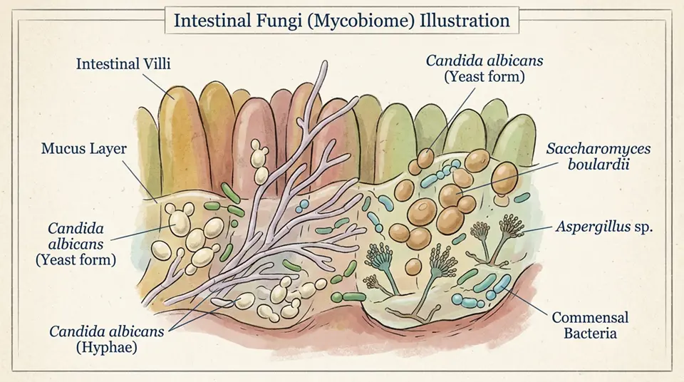
To understand the gravity of this situation, we have to understand the antagonist. If this were a crime novel, the culprit would be a sophisticated, stealthy assassin. In biology, it is the Mucorales order of fungi.
These molds are ubiquitous. They are the janitors of the natural world. Walk into a forest in the Pacific Northwest, kick over a pile of damp autumn leaves, or dig your hands into the soil, and you are surrounded by them. They thrive on decaying organic matter—compost, rotting wood, leaves.

Source: Wikimedia Commons, CC BY-SA 3.0
When Mucormycosis takes hold, it is aggressive. It affects the sinuses, the brain, or the lungs. It invades blood vessels, cutting off blood supply to tissue, causing that tissue to die and turn black—hence the terrifying moniker it sometimes carries, “Black Fungus.”
The spores are airborne. We breathe them in every day. You, reading this right now, have likely inhaled fungal spores within the last hour.

Source: Wikimedia Commons, CC BY-SA 3.0
Harborview has stated they are investigating. This likely involves industrial hygienists swabbing air ducts, testing linen supplies, checking water filtration systems, and analyzing the airflow pressure in patient rooms. They are looking for the “reservoir”—the hiding spot where the mold is growing and releasing its spores.
The Human Cost: Beyond the Statistics
While we analyze the science, we must pivot back to the “human core.” We are talking about six families.
Imagine the scenario: Your loved one is in the hospital. You are worried about their heart surgery or their trauma recovery. You trust the doctors. You trust the clean white sheets. Then, you are told that they have contracted a rare fungal infection.
A Critical Perspective: The YMYL Implications
From a Your Money Your Life perspective, this news is critical. Healthcare decisions are high-stakes. When a major regional hospital reports an outbreak, it affects public behavior.
The Paradox of the “Superbug” Era
We often focus heavily on antibiotic-resistant bacteria such as MRSA. Fungal infections are the silent cousins in this family of threats.
Conclusion: The Path Forward
The immediate next steps for Harborview are clear: isolate the source, sterilize the environment, and transparently communicate with the public.
References
Harborview Medical Center — Wikipedia
According to FOX13 SEATTLE
Key Takeaways
- Mucormycosis is a rapidly progressive and often fatal invasive fungal infection caused by Mucorales fungi, primarily affecting immunocompromised patients and those with poorly controlled diabetes mellitus.
- Hospital outbreaks of mucormycosis are associated with contaminated linen, cardboard boxes, construction dust, and HVAC system breaches that introduce Mucorales spores into clinical environments.
- The hallmark pathological feature of mucormycosis is angioinvasion—fungal hyphae penetrate and destroy blood vessel walls, causing thrombosis, infarction, and extensive tissue necrosis that drives the high mortality rate.
- Liposomal amphotericin B is the first-line treatment for most mucormycosis cases, often requiring extended high-dose courses alongside aggressive surgical debridement of infected tissue.
- Isavuconazole has emerged as an alternative or salvage treatment for mucormycosis, offering the advantage of oral bioavailability compared to intravenous amphotericin B formulations.
Frequently Asked Questions
What is mucormycosis and how is it different from other fungal infections?
Mucormycosis (formerly called zygomycosis) is a severe and rapidly progressive invasive fungal infection caused by fungi in the order Mucorales, which includes Rhizopus, Mucor, Cunninghamella, Lichtheimia (formerly Absidia), Apophysomyces, and other genera. It is distinct from other major invasive fungal infections in several clinically important ways. Distinguished from aspergillosis: Mucorales are phylogenetically unrelated to Aspergillus; Mucorales have broad, ribbon-like, aseptate (non-divided) hyphae, compared to Aspergillus’s septate hyphae with dichotomous (45°) branching; this morphological difference is visible on tissue microscopy and helps distinguish them. Mucorales are resistant to voriconazole (the preferred drug for aspergillosis)—a critical point, because empirical voriconazole for presumed aspergillosis will have no effect on mucormycosis and delays appropriate treatment; liposomal amphotericin B is the first-line drug for mucormycosis. The hallmark of mucormycosis: angioinvasion—Mucorales hyphae invade blood vessel walls with a predilection for arteries; vessel wall invasion leads to thrombosis (clotting), vessel occlusion, and infarction (tissue death from lack of blood supply); this is why mucormycosis produces extensive black necrotic tissue (eschar); the vascular destruction also limits drug delivery to infected tissue. Speed of progression: mucormycosis progresses from initial spore germination to extensive tissue destruction within days—faster than most other invasive fungal infections; this speed demands urgent diagnosis and treatment.
How do mucormycosis outbreaks happen in hospitals?
Hospital-acquired (nosocomial) mucormycosis outbreaks have been documented from multiple healthcare facilities globally, with the common thread being a contaminated environmental source that generates Mucorales spores in areas where susceptible patients are present. Documented outbreak sources: contaminated linen—multiple outbreak investigations have traced mucormycosis clusters to Mucorales-contaminated hospital linen (bedding, gowns), particularly when linen was stored in cardboard boxes that allowed moisture and hence fungal growth; a major outbreak at Duke University Medical Center in 2009 was traced to contaminated linen. Hospital construction and renovation—construction and renovation activities release large quantities of fungal spores from disturbed soil and building materials; Mucorales spores are present in construction dust; hospital construction adjacent to or within buildings where immunocompromised patients are cared for has been associated with multiple mucormycosis outbreaks; negative pressure rooms and air filtration barriers during construction are essential preventive measures. Contaminated medical supplies—adhesive tape (a contaminated batch caused cutaneous mucormycosis in neonatal ICUs), tongue depressors, and other medical supplies have been implicated. HVAC system defects—malfunctioning HVAC systems that failed to maintain positive pressure in protected haematology units allowed ingress of outdoor fungal spores; a filter failure or incorrect pressure differential can cause spore counts in haematology units to rise dramatically. Harborview Medical Center investigation: the specific outbreak at Harborview Medical Center (referenced in the article subject) involved investigating a cluster of mucormycosis cases for a common environmental source; these investigations require detailed epidemiological analysis comparing exposure patterns of cases versus non-cases to identify the likely source.
Who is most at risk for mucormycosis?
Mucormycosis has a very specific host risk profile, with certain medical conditions creating extreme susceptibility while healthy individuals with intact immune systems are effectively not at risk from environmental spore exposure. Highest-risk conditions: uncontrolled diabetes mellitus (particularly with diabetic ketoacidosis)—diabetic ketoacidosis creates an environment of elevated glucose, reduced serum pH, and iron availability that dramatically promotes Mucorales growth; neutrophils from diabetic patients show impaired fungal killing; mucormycosis in diabetic ketoacidosis typically presents as rhinocerebral (nose and sinus extending to brain) disease. Haematological malignancy with prolonged neutropenia—leukaemia and lymphoma patients receiving intensive chemotherapy develop severe neutropenia (very low neutrophil counts) during which Mucorales spores, if inhaled, cannot be destroyed; pulmonary mucormycosis predominates in this population. Stem cell transplant (HSCT) recipients—during engraftment and graft-versus-host disease treatment; both early and late post-transplant periods carry risk. Solid organ transplant recipients—particularly those requiring augmented immunosuppression for rejection episodes. Iron overload and deferoxamine therapy—iron is essential for Mucorales growth; iron overload and paradoxically, deferoxamine (an iron chelating drug used to treat iron overload) increases mucormycosis risk because deferoxamine-iron complexes are directly utilised by Mucorales as an iron source. COVID-19 associated mucormycosis (CAM)—became widely recognised during the Indian Delta COVID-19 wave in 2021; dexamethasone use for severe COVID, pre-existing uncontrolled diabetes, and possible zinc supplementation interacted to create a massive outbreak of rhinocerebral mucormycosis (‘black fungus’) in India.
What is the survival rate for mucormycosis?
Mucormycosis has among the highest mortality rates of any invasive fungal infection, with outcomes varying significantly by clinical form, anatomical site, and timeliness of treatment. Overall mortality rates by clinical form: rhinocerebral mucormycosis (sinuses + brain involvement) in diabetics—overall mortality approximately 25–50%; intracranial extension dramatically worsens prognosis (approaching 90% mortality); early aggressive surgical debridement of sinuses significantly improves survival. Pulmonary mucormycosis in haematological malignancy patients—overall mortality 50–75%; disseminated pulmonary mucormycosis approaches 90% mortality; less favourable outcomes than rhinocerebral form because surgical resection of lung tissue is less often feasible. Disseminated mucormycosis (involving multiple organs including brain)—mortality approaches 90–100%; survival is exceptional and typically only seen with very aggressive combined surgical and antifungal therapy. Cutaneous mucormycosis (in trauma or burns)—better prognosis with aggressive local debridement; mortality 20–30% in well-managed cases. Overall across all forms—reported mortality ranges from 40–80% in most case series; improving over time with better drugs and earlier diagnosis but still extremely high. Factors associated with better outcomes: early antifungal therapy (< 6 days from diagnosis); surgical debridement when anatomically feasible; control of underlying predisposing condition (ketoacidosis correction, immunosuppression reduction when possible); use of appropriate antifungal (liposomal amphotericin B rather than voriconazole). The most important prognostic factor: time to diagnosis and appropriate treatment—each week of delayed appropriate therapy dramatically worsens outcomes; the time-sensitivity of mucormycosis management rivals that of any invasive infection.
How is mucormycosis diagnosed and treated?
Mucormycosis diagnosis and treatment represent major challenges because the infection is rare (so clinical awareness is low), diagnosis requires invasive procedures, and effective treatment requires high-dose toxic drugs combined with extensive surgery. Diagnosis: tissue biopsy is the gold standard—tissue must be obtained from the infected site (sinus biopsy, bronchoscopy with BAL and endobronchial biopsy, surgical tissue) and examined by experienced pathologists; the characteristic broad, ribbon-like, aseptate hyphae with irregular branching on tissue microscopy is diagnostic; Mucorales grow on standard fungal culture media within 2–3 days (faster than Aspergillus). CT imaging—CT scan is essential for assessing the extent of infection in rhinocerebral form (evaluating sinus, orbit, and intracranial involvement) and pulmonary form; the ‘reverse halo sign’ on CT is associated with mucormycosis (area of ground glass opacity surrounded by ring of consolidation) but not specific. Serum beta-D-glucan—Mucorales are unusual among invasive fungi in NOT producing beta-D-glucan; a negative BDG test in a patient with risk factors and clinical features of invasive fungal infection raises the suspicion of mucormycosis specifically. PCR tests—molecular diagnostic tests for Mucorales DNA are under development and available in some specialist centres; not yet standardised or widely available. Treatment: liposomal amphotericin B 5–10 mg/kg/day IV—first-line drug; higher doses used than for aspergillosis; nephrotoxicity is the main dose-limiting side effect. Surgical debridement—essential for rhinocerebral and cutaneous forms; often requires repeated radical surgery. Isavuconazole—alternative or salvage therapy; can be used orally; has activity against Mucorales unlike other azoles. Posaconazole—used for continuation therapy after initial amphotericin B response. Iron chelation—deferasirox (not deferoxamine) has been studied as adjunctive therapy.