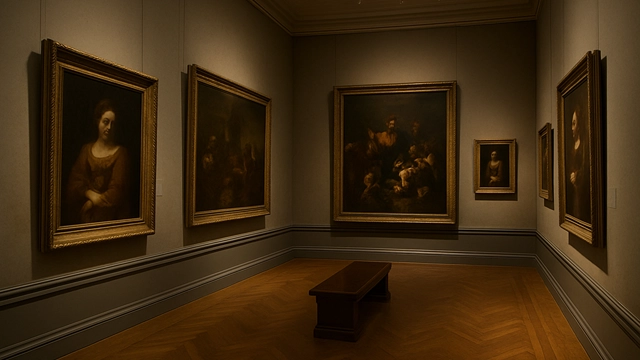
When we move through a museum gallery, we tend to notice the surface details: the warmth of pigment, the rhythm of brushstrokes, the patient glow of varnish. Yet in these quiet rooms, another kind of viewer is busy making judgments—mold. It behaves like a silent, particular, and slightly presumptuous aesthetic critic, drawn to the most vulnerable parts of an artwork, as if compelled to add its own marks to the masterpiece.

Its motives are simple. Give it moisture, darkness, and a hint of nourishment, and it begins its private project. Canvas fibers, wooden stretchers, protein-rich adhesives, the lipids in oil-based binders—these are all fair game. To mold, a painting is not a cultural treasure but a well-stocked buffet.
And this buffet has consumed more masterpieces than most visitors ever realize.
On Leonardo’s Last Supper, conservators have documented the effects not only of time and pollution but also mold—thin brown and black specks infiltrating the fragile paint layers. In the Roman catacombs, persistent humidity allows species such as Fusarium, Cladosporium, and Penicillium to drift across frescoes like a soft, unwanted mist. Even the famous Lascaux cave paintings once faced a severe fungal outbreak, forcing strict limits on human visits to avoid adding more moisture to an already delicate environment.

In these moments, mold behaves not simply as a destroyer but as an uninvited collaborator, altering surfaces in ways that echo deliberate artistic gestures.
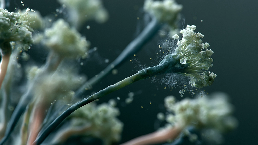
Under the microscope, its traces reveal a strange kind of beauty: networks of fine white filaments, powdery green blooms, mottled brown spots that mimic natural patina. Some colonies form threads so delicate they resemble intentional fog-like glazes; others produce granular textures difficult to distinguish from the canvas’s original tooth. As some conservators like to say, “Mold can be surprisingly meticulous—especially when destroying something.”
Behind this “handwriting” lies material science. Canvas made from cotton or linen provides abundant cellulose. Traditional grounds and adhesives based on animal glue supply proteins. Oil paint binders such as linseed oil contain fatty acids that certain fungi can digest. Even wooden frames can soften under the slow pressure of white-rot fungi. Mold’s interventions are not aesthetic decisions—they are biological necessities.

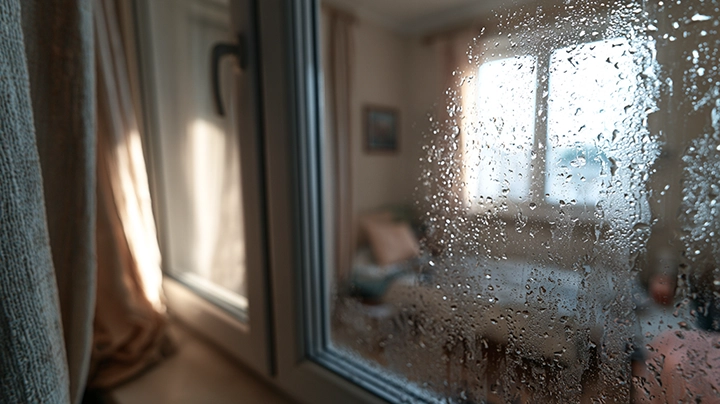
To restrain these uninvited artists, museums operate like slow-moving environmental laboratories. Relative humidity is kept near 50 percent, because once it climbs beyond 65 percent, fungal growth can accelerate dramatically. HVAC systems smooth out temperature swings that might trigger condensation. Paper-based collections are sometimes frozen to halt spores. Painted surfaces and frescoes undergo examinations with microscopes, molecular assays, or infrared and FTIR spectroscopy to track microbial activity.
Conservation teams deploy subtle countermeasures: volatile fungistatic compounds that suppress growth without disturbing pigment structures; gentle cleaning protocols that avoid damaging fragile surfaces; controlled darkness or cold rooms that give mold fewer chances to wake. Every step is part of a larger effort to keep mold from adding more strokes to the artwork.
Yet beneath this defensive choreography lies a quieter truth. Mold appears because artworks are built from materials that age—fibers, oils, gums, wood, paper. They breathe and absorb moisture. They shift and warp. And in those shifts, they invite microbes. Mold simply makes visible the fact that artworks, despite their cultural weight, are living negotiations with their environment.
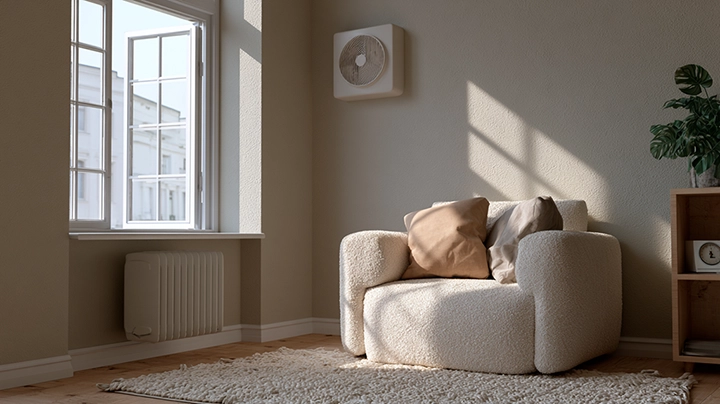
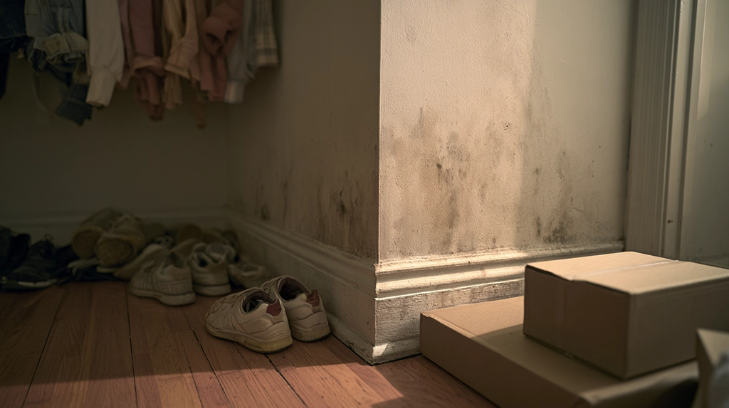
This negotiation happens far beyond museums: in family albums stored in dim corners, framed prints hanging on sunless walls, and small paintings sealed inside humid rooms. The same organisms at work in catacombs or caves can just as easily inhabit a bookshelf at home. Mold does not care about the prestige of its canvas; it follows only the logic of vulnerability.
So perhaps mold is not an aesthetic critic. It is a life form carrying out its ordinary habits, while we interpret its traces as commentary. The real question is how we read those traces—what they reveal about time, fragility, and the materials we trust to hold our memories.
When we see specks on an old painting, we might try thinking of them not merely as damage but as marginal notes written between an artwork, its environment, and the long, humid breath of history. Conservation is not about erasing these notes, but about keeping mold from writing new ones.
REFERENCES
Academic Sources
- Sterflinger, K. (2010). Fungi: Their role in deterioration of cultural heritage. Fungal Biology Reviews. https://doi.org/10.1016/j.fbr.2010.03.004
- Caneva, G., Nugari, M., & Salvadori, O. (2008). Biology in the Conservation of Works of Art. ICCROM.
- Dupont, A.‐L. (2020). Microorganisms and the biodeterioration of heritage materials. International Biodeterioration & Biodegradation.
Official / Institutional
- ICCROM — International Centre for the Study of the Preservation and Restoration of Cultural Property
https://www.iccrom.org/ - Smithsonian Museum Conservation Institute
https://www.si.edu/mci - American Institute for Conservation (AIC)
https://www.culturalheritage.org/
Key Takeaways
- Some mold species produce pigments during growth that have been exploited by artists—historically and in contemporary practice—creating artwork that uses living or preserved biological processes as aesthetic media.
- The relationship between mold, decay, and art raises philosophical questions about impermanence, entropy, and the boundary between creation and destruction that have attracted conceptual artists for decades.
- Mold pigments—including the red and yellow pigments of Monascus purpureus (red yeast rice) and the melanins of Aspergillus niger—are studied both as natural colorants with food applications and as subjects of artistic investigation.
- Conservation science confronts the challenges of mold as both subject (in decay-based art) and threat (in the degradation of conventional artworks), developing protocols to stabilise biodegradable artworks while preventing unwanted mold damage.
- Art history is full of examples where mold has inadvertently become the aesthetic agent—foxing on old paper, verdigris on oxidising surfaces, and the patina of age that is partly biological.
Frequently Asked Questions
Have artists deliberately used mold in their artwork?
Yes—a growing body of contemporary art explicitly incorporates mold as a medium, subject, or collaborative creative agent, challenging conventional boundaries between biology and aesthetics. Notable historical and contemporary examples: Joseph Beuys, the influential German conceptual artist, incorporated organic decay processes (including mold) in materials-based works that explored themes of entropy and transformation. Anna Dumitriu (UK bioartist) has created extensive works incorporating mold cultures and microbial processes, including works responding to antibiotic resistance and human-microbe relationships. Olafur Eliasson’s work with natural processes includes decaying organic materials. Heather Barnett works with slime molds (which though not true fungi are often grouped with them) as research and art collaborators, creating time-lapse works of their maze-solving and network formation. Artists working with fungi specifically often collaborate with mycologists, using species selected for their visual characteristics—colour, texture, fruiting body form—as aesthetic material. These works frequently address themes of mortality, ecology, microbiome, and the invisibility of biological processes to human perception.
What pigments do molds naturally produce?
Mold pigment production is a well-studied area both for food industry applications and for understanding fungal secondary metabolism. The range of mold pigments is extraordinary: orange and red azaphilone pigments: Monascus purpureus and related species produce the red, orange, and yellow pigments of red yeast rice (hong qu in Chinese); these water-soluble pigments have been used as natural food colorants in East Asia for over a thousand years; they are being studied as natural alternatives to synthetic food dyes. Yellow and greenish pigments: Aspergillus species (A. flavus, A. parasiticus) produce aflatoxins (which are toxic) but also other yellow fluorescent pigments; various Penicillium species produce yellow compounds. Blue and green pigments: Penicillium species and some Fusarium produce blue-green pigments; Chlorociboria aeruginascens, a wood-rotting fungus, produces xylindein, a striking teal-blue compound that stains wood and has been investigated for art and textile applications. Black melanins: widely produced across many mold species, melanins are high-molecular-weight dark pigments in spore walls that confer UV resistance and stress tolerance. Red anthraquinone pigments: Cordyceps and other Hypocreales produce red anthraquinone compounds.
How do art conservators deal with mold damaging artworks?
Art conservation’s relationship with mold has two aspects: protecting artworks from unwanted mold damage (by far the larger concern) and, increasingly, stabilising artworks that deliberately incorporate living processes. Preventing mold damage to conventional artworks: the primary strategy is environmental control—maintaining museum and archive storage at 45–55% RH and 18–21°C (below the humidity threshold for most common art-damaging fungi such as Aspergillus, Penicillium, and Trichoderma); monitoring for elevated RH especially in basements, exterior walls, and flood-prone areas; using HEPA filtration to reduce airborne spore loads; using UV-blocking glazing and display cases to prevent photodegradation that can create substrates for mold. Treatment of mold-damaged objects: specialist paper and textile conservators use controlled low oxygen (argon or CO₂) anoxic treatment to kill mold on sensitive objects without chemical treatment; biocide treatment is controversial due to potential chemical damage to organic materials; dry brushing and HEPA vacuuming under controlled conditions; consultation with microbiologists for identification of species before treatment. Living or decaying artwork: ‘bioart’ presents unique conservation challenges—documenting the work’s intended appearance at various stages; decisions about whether and how to stabilise or arrest decay; preservation through documentation (photography, video, 3D scanning) rather than physical preservation.
What is the ‘foxing’ seen on old books and paper, and is it mold?
‘Foxing’ refers to the brownish spots or patches commonly seen on aged paper—books, photographs, maps, prints—that has been stored in imperfect conditions over years or decades. The origin and exact mechanism of foxing has been debated in conservation science for over 150 years, and research suggests it is multifactorial rather than having a single cause. Evidence for biological origin (mold): several studies have identified fungal hyphae, spores, and biological material within foxing spots using microscopy; DNA analysis has detected Aspergillus, Penicillium, and other species in foxed paper. Chemical evidence for mold: compounds characteristic of fungal metabolism (ergosterol, certain fatty acids) have been detected in foxing spots. Evidence for chemical (non-biological) origin: some foxing spots contain no detectable biological material; the pattern of foxing correlates with metal contamination (particularly iron and copper from manufacturing or water exposure) that can catalyse oxidative browning reactions independent of biology. Current scientific consensus: foxing is likely a complex phenomenon with both biological and chemical components that may interact—metal contamination may support mold growth; mold activity may catalyse chemical browning reactions; both can occur simultaneously. The relative importance of each mechanism varies between paper types, inks, and storage histories.
Why are some molds so visually striking?
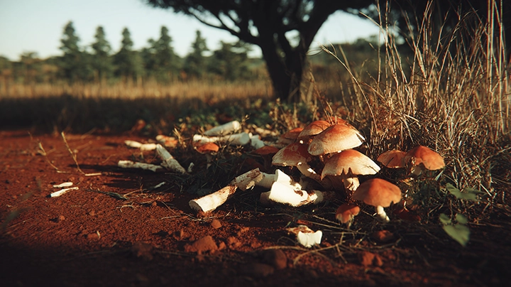
The visual characteristics of molds—their textures, colours, patterns, and three-dimensional structures—are products of evolutionary adaptation to biological functions rather than aesthetic selection, yet they produce remarkable visual effects that have attracted scientific and artistic attention. Spore colours: spore pigmentation serves protective functions—melanin in dark spores (Aspergillus niger’s black, Cladosporium’s olive green) provides UV protection that extends spore viability during atmospheric dispersal; carotenoid pigments in orange and yellow spores serve antioxidant functions. Spore colours are therefore concentrated in the spore coat where protection is needed, giving mold colonies visual patterns that change as sporulation proceeds. Colony morphology: the intricate radial symmetry of mold colonies growing on uniform media reflects the uniform hyphal extension rate in all directions from the inoculation point; variations in temperature, nutrient gradients, and competing organisms produce irregular patterns and differentiation. Fruiting body complexity: the macroscopic fruiting bodies of higher fungi (mushrooms, bracket fungi, cup fungi, stinkhorns) show extraordinary structural complexity and visual variety—these structures have evolved specifically for spore dispersal, with different architectures optimised for different dispersal mechanisms (gill spacing in agarics, tube density in boletes, cup shape in cup fungi). The visual drama of decay fungi—bright orange brackets on fallen logs, purple Cortinarius mushrooms, glowing bioluminescent species—reflects biochemical adaptations and evolutionary pressures that coincidentally create visual spectacle.