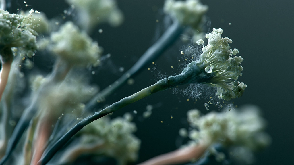
Invasive mold infections are among the deadliest complications faced by patients with weakened immune systems—especially those undergoing chemotherapy, organ transplantation, or immunosuppressive therapies. These infections are notoriously elusive, often going undiagnosed until irreversible damage has occurred. Now, a promising new imaging technology may offer clinicians a powerful new way to detect and monitor fungal diseases before it’s too late.
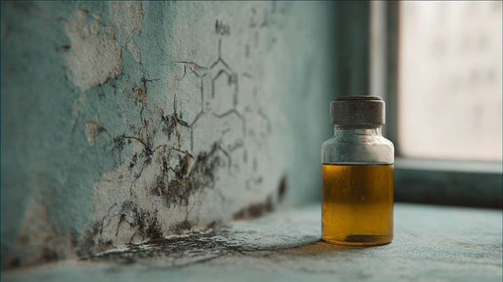
A team of researchers has unveiled a noninvasive diagnostic breakthrough: 18F-fluorodeoxysorbitol (18F-FDS)positron emission tomography (PET), a technique that allows fungal infections to be visualized in real time throughout the body. This approach could revolutionize how clinicians detect, differentiate, and treat mold-related diseases—particularly those caused by difficult-to-diagnose or drug-resistant fungi.

Visualizing the Invisible: A Diagnostic Leap Forward
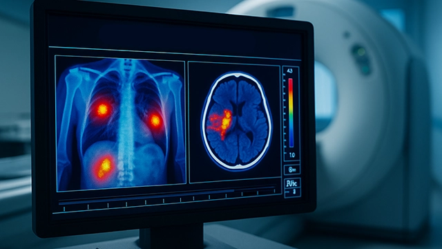
The study tested 18F-FDS PET in nine patients suspected of having invasive fungal infections. Four of those cases were later confirmed by traditional culture or molecular diagnostics. In each of the confirmed cases, the PET scans provided early, high-resolution images of mold lesions in the lungs and brain, including infections caused by azole-resistant strains and non-Aspergillus species typically missed by standard biomarker tests.
Unlike conventional diagnostics—which often rely on indirect indicators like galactomannan or β-D-glucan, and require invasive biopsies—18F-FDS PET directly visualizes fungal activity. It does so by exploiting a key biological difference: 18F-FDS accumulates inside fungal cells but not in human tissues, tumors, or sites of sterile inflammation.
A Tool for Precision Mycology
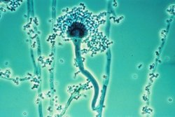
What makes this technique even more promising is its broad applicability. The tracer was shown to be selectively taken up by multiple pathogenic molds, including Aspergillus, Mucor, and Rhizopus. Animal models mirrored these human findings. Infected mice with lung or sinus fungal infections also showed precise tracer accumulation in the affected tissues.


Scalable, Accessible, and Ready for Clinics
One of the most important advantages of this technology is its scalability. The tracer, 18F-FDS, can be synthesized from 18F-FDG, a compound already used worldwide for cancer PET imaging. This means that most modern hospitals already possess the infrastructure and supply chain needed to produce and use this new tool.
Unlike other novel diagnostics that require specialized labs or training, 18F-FDS PET fits easily into existing imaging workflows. The scans are conducted within 2 to 3 hours after injection, delivering actionable insights in a timeframe that can change clinical decisions on the same day.

Real-Time Monitoring and Future Applications
Beyond initial detection, researchers are optimistic that 18F-FDS PET could be used to monitor antifungal treatment efficacy over time. If the tracer uptake diminishes during therapy, it could provide direct evidence of treatment success—a feature not currently available with traditional tests.
Looking forward, the team envisions mapping species-specific uptake patterns, creating an imaging-based library of fungal fingerprints. This could lead to tailored treatment plans, especially valuable in managing multidrug-resistant or mixed-species infections.
Hospitals could also integrate fungal PET scanning into infection control strategies for high-risk departments like hematology, oncology, and transplant units. The technology could help identify environmental outbreaks early and track the spread of airborne or systemic infections.
A Paradigm Shift in Fungal Diagnostics
Despite being in early clinical stages, the promise of 18F-FDS PET is undeniable. It addresses the core failures of current fungal diagnostics: invasiveness, species bias, and diagnostic delay. By offering a fast, sensitive, and noninvasive way to detect fungal infection system-wide, it could save lives where time is the most critical factor.
For decades, invasive mold infections have been a diagnostic black hole—clinically devastating yet largely invisible. This technology turns the lights on. For the first time, medicine can not only suspect a mold infection but see it.
As larger clinical trials move forward, experts hope to establish this method as a standard of care for immunocompromised patients. With fungal diseases on the rise globally due to climate change, such a tool could not come at a more urgent moment.
This innovation marks a pivotal moment in the war against invasive mold. From the metabolic uniqueness of fungi comes a visual signature that can now be tracked, measured, and responded to in real time. We are no longer groping in the dark for clues. We now have a direct visual link to our microbial adversaries.
The future of fungal diagnostics isn’t just about better tests. It’s about seeing fungal infections before they strike fatally. Thanks to 18F-FDS PET, that future may already be arriving—and for many patients, not a moment too soon.
References
- Ruiz-Gonzalez CE et al. (2025). 18F-FDS PET imaging for detection of invasive mold infections. Nature Communications. Article
- Lai J et al. (2021). Evaluation of 2-[18F]-Fluorodeoxysorbitol PET Imaging in Preclinical Models of Aspergillus Infection. Journal of Fungi. PMC
- ClinicalTrials.gov. NCT05611892 – 18F-FDS PET for Invasive Mold Infections. Registry
- IPCC. Climate Change Reports. IPCC
Key Takeaways
- PET (positron emission tomography) imaging combined with specific radioligands is being developed to visualise invasive fungal infections in living patients, enabling earlier and more precise diagnosis.
- Current diagnosis of invasive fungal infections relies on blood tests, cultures (which can take weeks and miss culture-negative disease), and CT imaging (which detects tissue damage rather than active infection).
- New PET tracers targeting fungal-specific cell wall components (such as siderophores—iron-scavenging molecules unique to certain fungi) can differentiate active fungal infection from bacterial infection or non-infectious inflammation.
- The ability to image fungal infection in real time could reduce the time from symptom onset to diagnosis from the current average of several days to hours, potentially saving lives.
- Early clinical studies of 68Ga-triacetylfusarinine C (68Ga-TAFC) PET imaging for Aspergillus infection have shown promising sensitivity and specificity, with ongoing trials.
Frequently Asked Questions
Why is diagnosing invasive fungal infections so difficult with current methods?
Diagnosing invasive fungal infections (IFIs) like invasive aspergillosis and mucormycosis is challenging for several reasons. Blood cultures are insensitive for many fungi (Aspergillus rarely produces positive blood cultures); bronchoscopy with bronchoalveolar lavage requires an invasive procedure and has variable sensitivity depending on operator skill and disease location; serum biomarkers like galactomannan and beta-D-glucan have useful sensitivity but can give false positives with certain antibiotics and foods; CT imaging shows characteristic radiological signs (halo sign, air crescent sign) but these appear after significant tissue damage has already occurred; and tissue biopsy is the gold standard but is often too risky in already coagulopathic, thrombocytopenic immunocompromised patients.
How does PET imaging work for detecting fungal infections?
Positron emission tomography (PET) imaging uses radioactively labelled molecules that are injected intravenously and accumulate in tissues with specific biological properties. Standard [18F]-FDG PET imaging detects increased glucose metabolism (present in both infection and tumour) but cannot distinguish fungal from bacterial infection or tumour. Fungal-specific PET tracers work differently: they exploit biological molecules that fungi uniquely require. Siderophores—small molecules secreted by Aspergillus to scavenge iron—can be labelled with gallium-68 (which competes with iron in the siderophore binding site). The labelled siderophore (68Ga-TAFC) is preferentially taken up by Aspergillus-infected tissue, producing a signal that specifically indicates the location and extent of active Aspergillus infection.
What is the clinical significance of being able to image fungal infections?
Imaging-guided diagnosis of fungal infections could transform clinical management in several ways: earlier diagnosis when infection burden is still manageable; ability to monitor treatment response in real time (seeing whether antifungal therapy is reducing infection burden); guidance for surgical resection of focal fungal lesions; differentiation of residual inactive infection from active disease at end of treatment; and potentially guiding antifungal therapy duration based on biological rather than arbitrary endpoint timing. For conditions like mucormycosis, where early surgical debridement combined with antifungal therapy can be life-saving but the extent of surgery needed is hard to judge, pre-operative imaging of infection boundaries could be particularly valuable.
Which fungal pathogens can currently be imaged with PET?
PET imaging research for fungal infections is most advanced for Aspergillus species, using siderophore-based tracers (68Ga-TAFC, 68Ga-DFOB). Mucormycosis presents a different challenge—Mucorales produce different siderophores—but 68Ga-labelled deferoxamine (which Mucorales species actively take up) is being explored. For Candida, which does not produce siderophores in mammalian hosts, alternative targeting strategies are under investigation. The challenge is that different fungal pathogens have different biology, requiring development of separate targeted tracers for each clinically significant species, substantially increasing the research and regulatory burden.
When might fungal-specific PET imaging become clinically available?
68Ga-TAFC PET imaging for invasive aspergillosis is in Phase 2 clinical trials in Europe and has shown strong performance characteristics in early studies. Regulatory approval timelines vary by jurisdiction—in the EU, novel PET tracers follow the radiopharmaceutical regulatory pathway; in the US, FDA clearance requires Phase 3 clinical trial data demonstrating clinical utility. Optimistic projections suggest that fungal-specific PET imaging could be available in specialist centres (academic medical centres with established PET programmes) within 5–10 years for Aspergillus, with broader availability thereafter. Cost and access to PET scanners will be limiting factors for widespread uptake in lower-resource settings.